Impacted Wisdom Teeth Touching the Nerve — How CBCT Changed the Conversation
A panoramic X-ray showed deeply impacted lower wisdom teeth dangerously close to the mandibular nerve. But it was the 3D view from the CS 8200 that gave this clinician the diagnostic confidence to plan extraction safely — and the images to explain the risks to his patient.
 Dr Hubertus Schindler
A 30-year-old male presented with recurrent pain from his lower right wisdom tooth. Initial examination revealed a complicated picture: both lower wisdom teeth (LR8 and LL8) were extremely deeply impacted, with signs of coronal osteolysis around the right side causing the pain. On top of this, the patient had poor oral hygiene resulting in gingivitis and calculus build-up, caries in multiple teeth, and wear patterns consistent with bruxism.
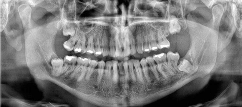
The panoramic radiograph immediately raised the critical concern: both impacted wisdom teeth appeared to be in direct contact with the inferior alveolar nerve (IAN) canals. On a 2D panoramic, the relationship between an impacted tooth root and the nerve canal is often ambiguous — the structures overlap, and you cannot tell whether the root is buccal, lingual, or truly in contact with the canal. This is the exact scenario where extraction carries the highest risk of nerve injury.
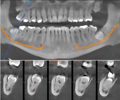
A CBCT scan was acquired using the Carestream CS 8200 to determine the exact 3D relationship between the impacted roots and the IAN canals. The difference between the panoramic view and the CBCT was immediate and clinically decisive.
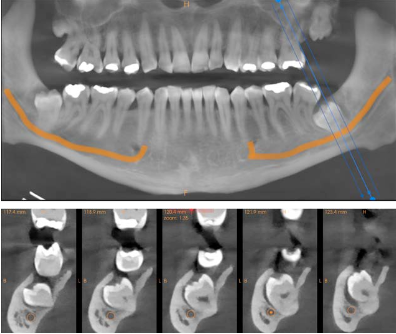
Cross-sectional views of both sides showed the precise spatial relationship that the panoramic could only hint at. On the right side (LR8), the CBCT confirmed direct contact between the root and the nerve canal. On the left (LL8), the tooth was in close proximity but not in direct contact — a distinction that significantly affects the surgical approach and the informed consent conversation.
But the most powerful output was the 3D volume rendering. The CS 8200's software generated fully rotatable 3D views of both impacted teeth and their nerve canals, showing the anatomical relationship from any angle. These images did not just help the clinician plan — they transformed the patient conversation.
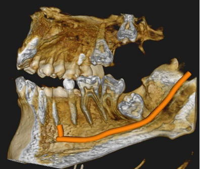
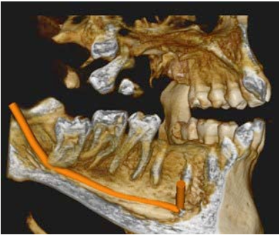
The images captured so much detail that they helped the patient understand his condition and why the proposed treatment was necessary. This improved the quality of his informed consent. I had all the information I needed, and gaining treatment acceptance was straightforward because the patient was properly informed.
— Dr Hubertus SchindlerWith the full diagnostic picture from the CS 8200, a comprehensive treatment plan was developed and presented to the patient using the CBCT images on screen in CS Imaging software — all in the same appointment, with no need to refer out for 3D imaging or schedule a separate consultation.
The patient accepted all four treatment recommendations. Dr Schindler noted that the CS 8200 eliminated the need for a separate referral for CBCT — the scan was taken and reviewed in the same session, streamlining the diagnostic workflow and allowing the full treatment plan to be discussed immediately.
A panoramic is not enough when wisdom teeth are near the IAN canal. When a 2D image shows the root overlapping the nerve, you cannot determine the true spatial relationship. CBCT cross-sections and 3D rendering give you the exact answer — is the root buccal, lingual, or in direct contact? That distinction determines the surgical approach and the risk you need to communicate.
3D images are a consent tool, not just a diagnostic tool. Showing a patient a volume-rendered view of their impacted tooth wrapped around the nerve canal is worth a thousand words. It transforms informed consent from a verbal exercise into a visual one — and patients who truly understand the risks give better quality consent.
In-house CBCT eliminates the referral bottleneck. Dr Schindler previously referred out for 3D imaging, which delayed diagnosis and required an extra appointment. With the CS 8200 in practice, the scan, review, and treatment discussion all happen in the same visit — better patient experience, faster case acceptance.
You do not need to be a specialist to use CBCT effectively. The CS 8200 was Dr Schindler's first CBCT system. He found it easy to learn and intuitive in daily practice. But reading a CBCT well is a skill that develops with training — our CBCT Level 2 Reporting Course builds the systematic interpretation skills that let you get the most from every scan you take.
Carestream CS 8200
Our most popular CBCT system. The CS 8200 combines panoramic, cephalometric, and 3D imaging in a single unit with an intuitive interface and fast acquisition. Multiple field-of-view options from focused 5×5 cm scans to full jaw coverage. Ideal for general practice, oral surgery, and implantology.
View CS 8200 specifications →Thinking about bringing CBCT in-house? The CS 8200 is our most popular system for a reason.