A Driving Instructor Who Couldn't Drive — How Digital Dentistry Treated Her Sleep Apnoea
A 53-year-old professional driving instructor diagnosed with severe OSA — unfit to drive, career at risk. A fully digital workflow using panoramic imaging and intraoral scanning delivered a mandibular advancement device that brought her AHI score from 34 down to 2.6. Life-changing dentistry.
 Dr Paul Reaney
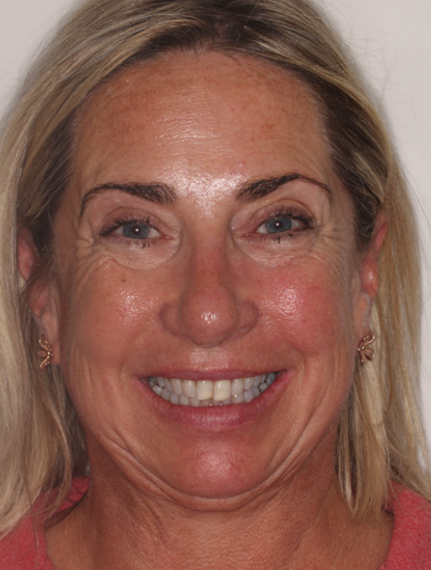
A 53-year-old woman was referred by her GP following a hospital sleep study. She had suffered a stroke before the age of 50 and was experiencing extreme fatigue affecting her quality of life. The sleep study confirmed the cause: an Apnoea/Hypopnoea Index (AHI) score of 34 — consistent with severe Obstructive Sleep Apnoea.
The diagnosis had immediate consequences beyond her health. With an AHI of 34, she was legally unfit to drive. For a professional driving instructor, this was not just a health concern — it was career-ending news.
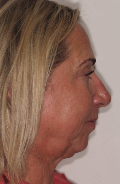
Clinical evaluation found a moderate Class II skeletal relationship with a retrognathic mandible. The lack of space for the tongue was contributing to airway collapse during sleep. Assessment followed British Society of Dental Sleep Medicine guidelines, including checks for sleep-related bruxism, dental erosion, and soft tissue indicators such as scalloped tongue.
The patient was sent home with a NightOwl home sleep monitor for baseline screening and later titration of the oral appliance. Based on the clinical findings and sleep data, it was decided that a fully digital CAD/CAM workflow would deliver the best outcome — a smaller, better-tolerated ResMed Narval CC Mandibular Advancement Device.
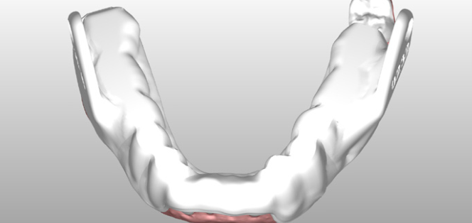
Digital impressions were captured using an intraoral scanner with CS 3600 acquisition software. This was critical because mandibular advancement devices require multiple bite registrations at different protrusion levels — something that digital scanning handles far more accurately than traditional impressions.
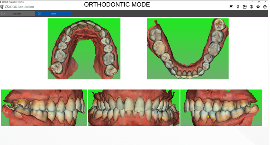
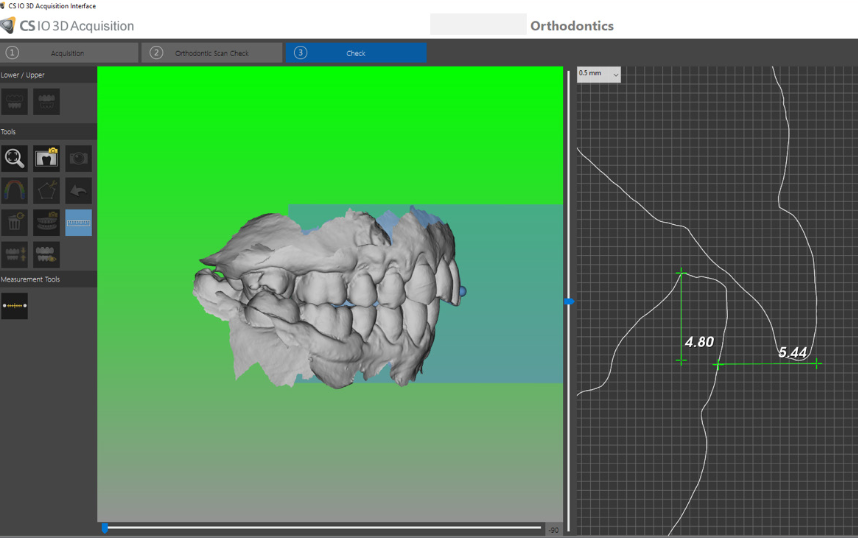
The software's undercut tools helped identify where tooth anatomy could be modified — for example, using direct composite attachments to create undercuts that improve appliance retention. Overjet and overbite measurements were recorded at the initial scan for comparison at future reviews to monitor any unwanted tooth movement.
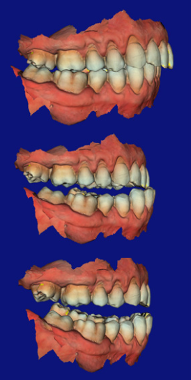
The full arch scans were exported to ResMed, where the 3D model was aligned with a virtual articulator and the Narval CC appliance designed using a completely digital workflow — no physical models, no alginate, no stone.
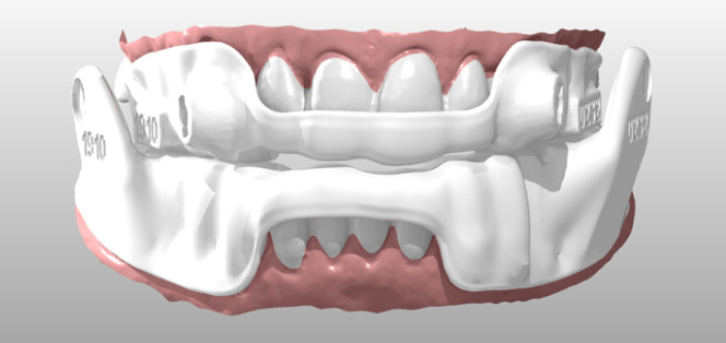
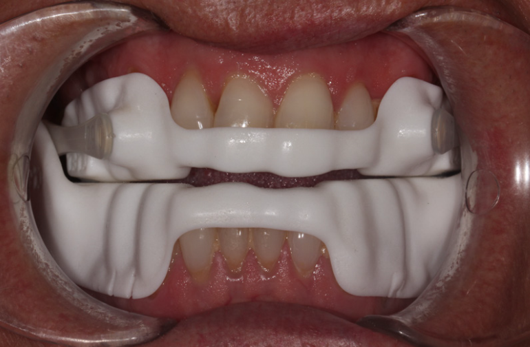
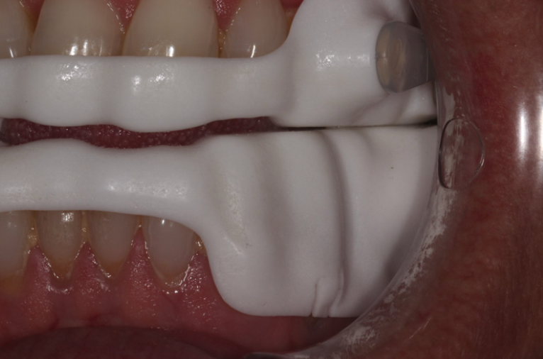
On fitting day, the device was checked for retention, occlusion, TMJ comfort, connector mechanics, and protrusion accuracy. The fit was accurate and comfortable — reinforcing the benefits of a fully digital impression and design workflow over traditional methods.
The result was transformative. Post-treatment sleep monitoring showed:
An AHI below 5 is considered normal. This patient went from severe sleep apnoea — unable to drive, extreme fatigue, elevated stroke risk — to a normal sleep score. She got her driving licence back. She got her career back. She got her life back.
For this patient, dental sleep medicine gave her back her livelihood and her quality of living. As dentists look for opportunities to expand their treatment options, combining a fully digital workflow with the right sleep lab or partner can help them change the lives of countless patients.
— Dr Paul ReaneyDental sleep medicine is a genuine practice builder. OSA affects an estimated 1.5 million adults in the UK. Many are undiagnosed or poorly managed. Mandibular advancement devices are a proven treatment — and dentists are uniquely placed to provide them. This is a clinical service that changes lives and generates recurring revenue.
A digital workflow produces better-fitting, better-tolerated appliances. Digital scanning captures multiple bite registrations accurately, eliminates the distortion risks of alginate, and enables a fully CAD/CAM-designed device that is smaller and more comfortable than traditionally made alternatives.
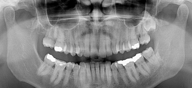
Panoramic imaging is essential baseline assessment. The CS 8100 panoramic provided the full picture — root morphology, periodontal status, TMJ assessment, and detection of any undiagnosed pathology — before appliance therapy began. You cannot safely fit an MAD without this information.
The equipment you already own may be all you need. If you have a Carestream panoramic system and an intraoral scanner, you already have the digital capture tools for this workflow. The clinical knowledge is the missing piece — and that is learnable. Talk to us about how to get started with dental sleep medicine in your practice.
Carestream CS 8100
Extraoral panoramic imaging system providing the essential baseline assessment — root morphology, periodontal status, TMJ, and pathology screening before appliance therapy.
View CS 8200 →Intraoral Scanner
Digital impression capture with CS 3600 acquisition software. Multiple bite registrations, undercut analysis, and accurate protrusion recording for MAD design.
Compare intraoral scanners →Interested in adding dental sleep medicine to your practice?