AI-Assisted Implant Planning: Crown First, Implant Second
A missing upper left first molar, a sinus close by, and a patient who had already seen what happens when a gap is left alone. Dr Kunal Shah planned the crown first and let the software propose the implant.
 Dr Kunal Shah
A 40-year-old man came in with discomfort when chewing on his upper left side. He was medically fit and well. The upper left first molar (UL6) was missing. Food was packing into the space, and he found the area hard to keep clean.
He had a clear reason to act. More than ten years earlier he lost his lower left second premolar, and the first molar behind it drifted into the gap. He did not want the same thing to happen again, or for the opposing tooth to over-erupt.
Every option was discussed: no treatment, a removable prosthesis, a fixed bridge, and an implant. He already had an implant in his upper right quadrant, so he knew the process. He chose an implant.
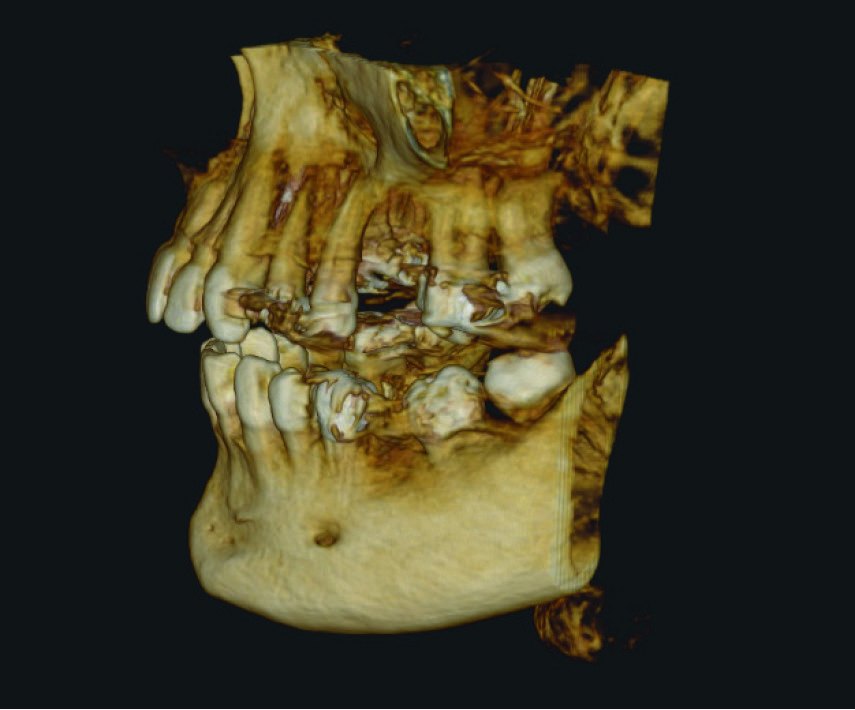
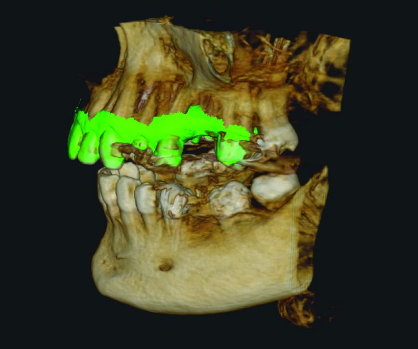
Clinical and radiographic assessment confirmed enough bone for an implant, with good periodontal health and nothing to rule implant treatment out. The anatomy to respect was the maxillary sinus, which sat close to the planned site.
An 8 x 9 volume was taken on a Carestream Dental CBCT. An intraoral scan was then imported and matched to it in CS 3D Imaging Premium.
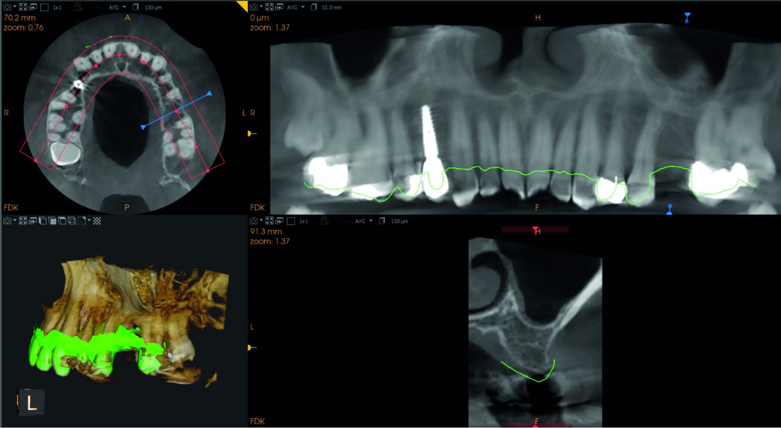
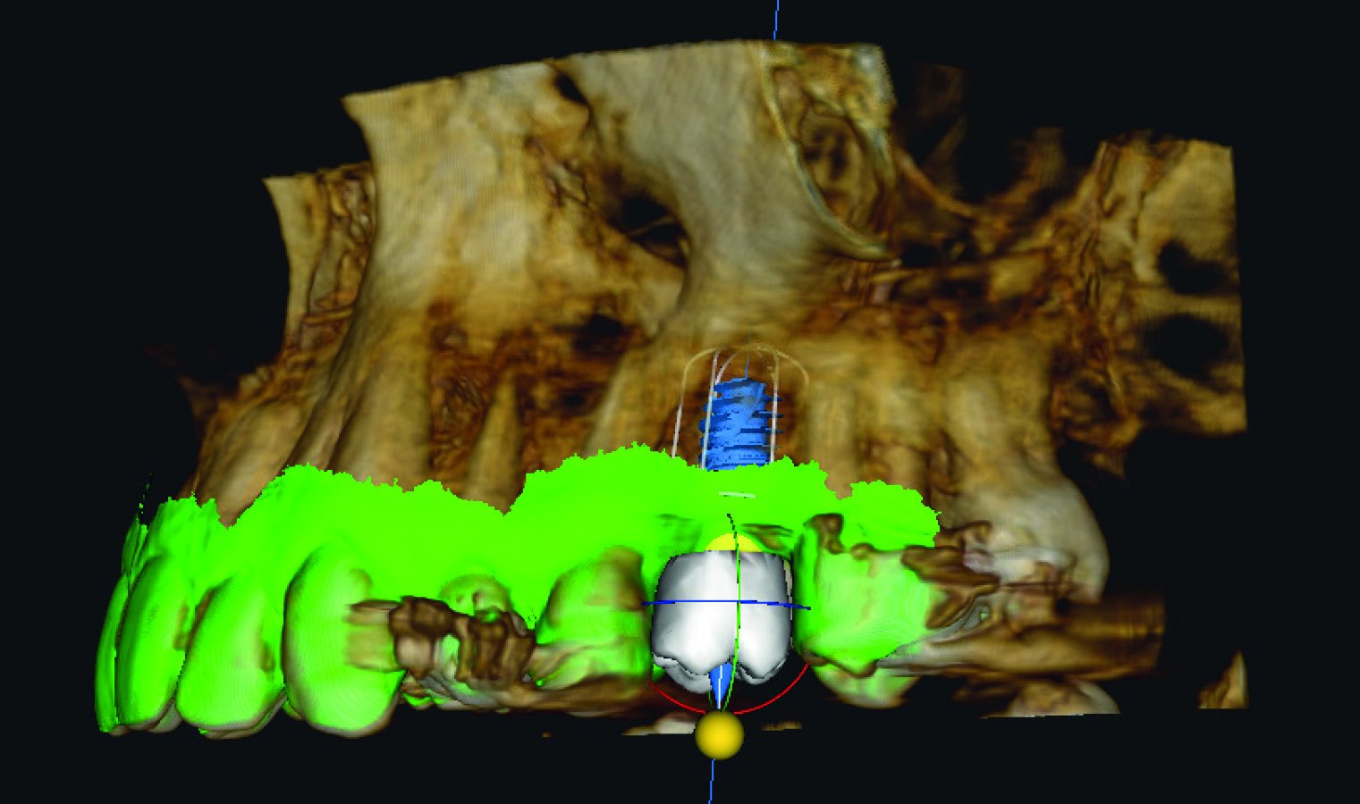
Planning was crown first. The ideal crown was set before any implant was placed on screen. The software's AI-assisted auto implant planning then proposed an implant position based on that crown, the surrounding anatomy and the bone available.
Dr Shah checked the proposal against the things that matter in the posterior maxilla: the distance to the sinus, the bucco-palatal width of the ridge, and the angulation.
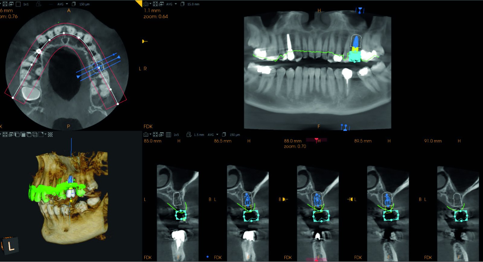
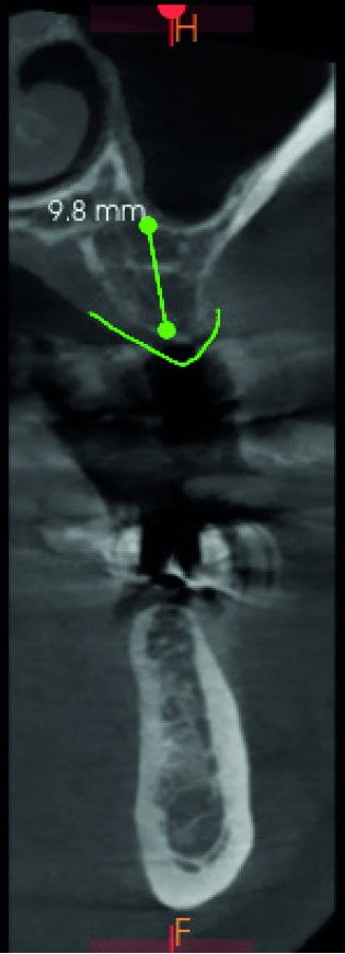
The digital plan could have been turned into a surgical guide. In this case Dr Shah chose a conventional freehand approach, with the plan as his reference. The implant was an Anthogyr Axiom X3, chosen for its suitability in the posterior maxilla.
The implant was placed successfully in the UL6 region.
AI-generated proposals are an adjunct, not a replacement for clinical judgement. The clinician stays fully responsible for checking the proposed plan and changing it where needed.
— Based on the clinical findings of Dr Kunal ShahWant to learn implant planning hands-on?
The 360 Implant Academy is two hands-on days at Octagon House in Yorkshire. Plan a real case, print your own surgical guide, then place an implant on the model you take home. 16 delegates, 12+ CPD hours, £495 + VAT.
See the 360 Implant Academy →Plan the crown, then the implant. Setting the restoration first means the implant goes where the tooth needs it, not only where the bone is easiest.
AI planning saves time at the planning stage. Dr Shah reports reduced planning time and simpler data handling, with the CBCT and intraoral scan matched in one place.
You can see the risk before you pick up a drill. Bone dimensions, crown position and the distance to the sinus are all visible on one plan.
The software proposes. You decide. A sound grasp of surgical anatomy, prosthetic principles and conventional implant workflows is still what makes the outcome predictable.
CS 3D Imaging Premium
Matches your CBCT and intraoral scan, sets the crown and proposes the implant position with AI. 360 Visualise supplies, installs and supports it alongside the Carestream CBCT range.
See CS 3D Imaging Premium →Want to plan implants like this in your own practice?